Hypertensive retinopathy is a slowly developing condition where high blood pressure damages blood vessels in the retina. The retina is the lining at the back of each eye that receives light from objects and turns it into signals, which are transported to the brain for interpretation. This enables us to understand what we see.
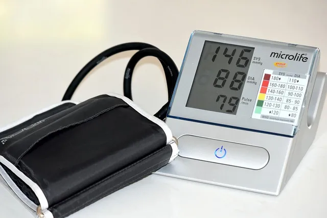
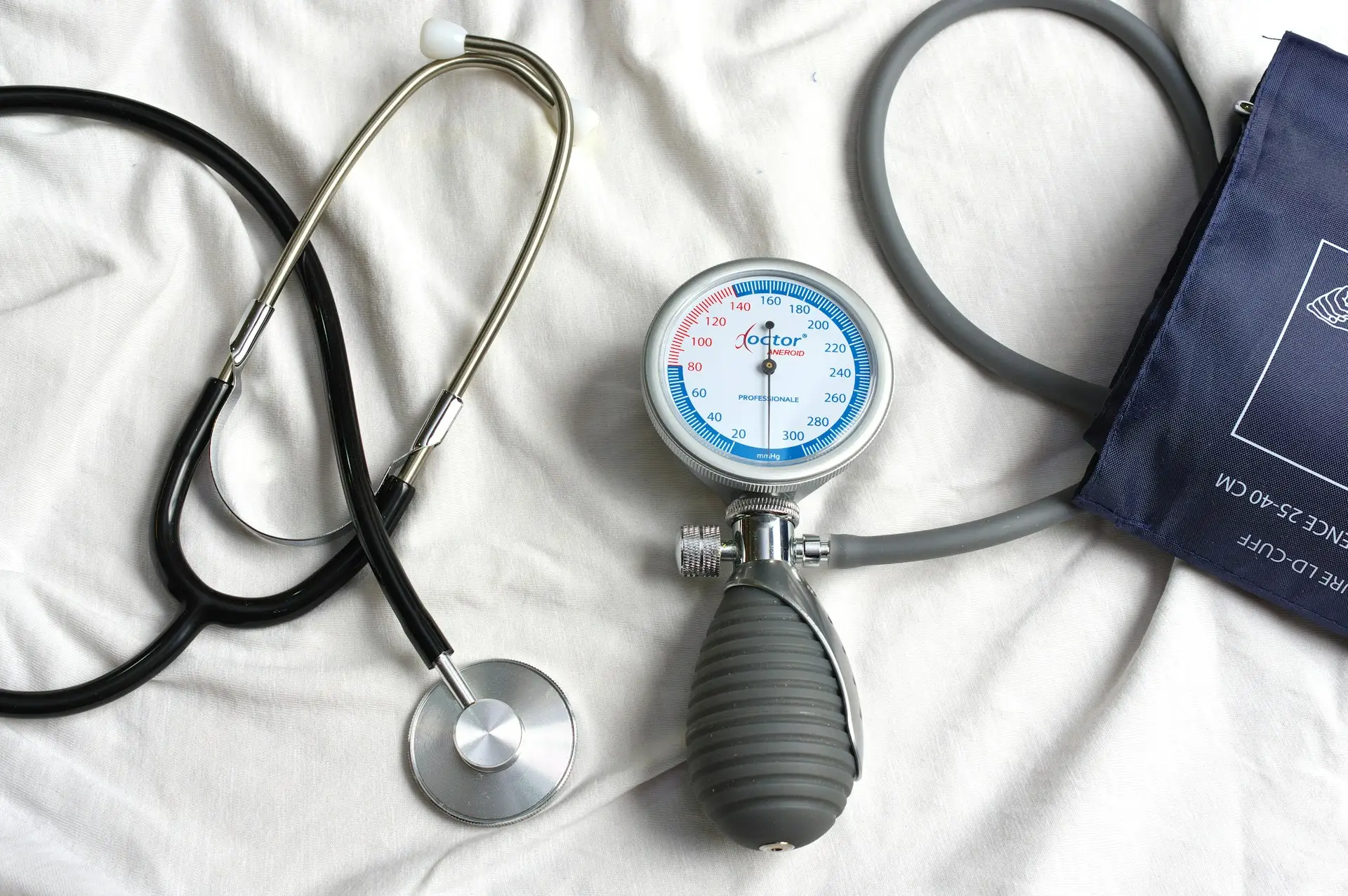
High blood pressure (hypertension) means that the force your blood exerts on the walls of your arteries is higher than normal.
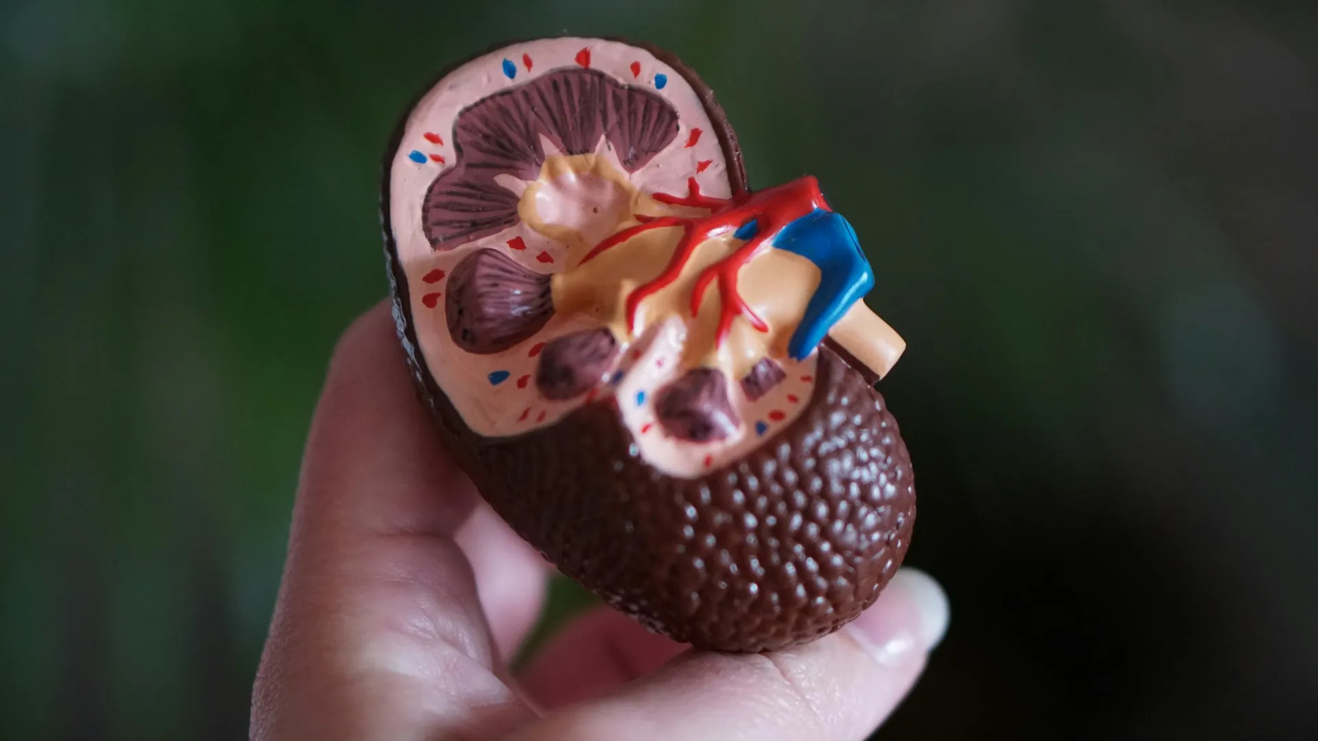
Over time, high blood pressure affects blood vessels throughout the body and can cause different organs and tissues to malfunction, including the eyes. Actually, hypertensive retinopathy may be one of the early signs that high blood pressure is damaging many other organs, including vital ones like the heart, kidneys, and brain.
Hypertensive retinopathy affects about 4% to 18.7% of hypertensive people who don’t have diabetes. It affects men more often than it affects women.
How does high blood pressure damage the retina?
Initially, high blood pressure causes the blood vessels in your retina to tighten and become narrow (vasospasm). This can occur at one point in the blood vessel, or throughout its length. This reduces the blood flow to your retina.
With time, the high blood pressure causes the walls of the blood vessels to become thick and inelastic. This further worsens blood flow and oxygen supply to the retina.
Seriously high blood pressure can also damage the blood vessel walls and cause blood to leak into the different parts of the retina, causing visual disturbances or vision loss.
What are the symptoms of hypertensive retinopathy?
Most people usually have no symptoms until the condition is severe. If you have high blood pressure, it is important to seek prompt medical help when you develop any problems with your vision. Other symptoms may include headaches and eye pain.
Who is at risk of developing hypertensive retinopathy?
Not everyone with hypertension will have their retina affected. You may have higher chances of getting hypertensive retinopathy if you:
- Have untreated high blood pressure for a long time
- Have certain genes
- Smoke
- Have kidney dysfunction
- Are Afro-Caribbean or of Chinese descent
- Have high blood levels of cholesterol
How is hypertensive retinopathy diagnosed?
Diagnosing hypertensive retinopathy is a multistep process where an eye doctor will:
- Take a medical history. You will be asked questions about any symptoms, how long you have had hypertension, what medications you are taking and how well your blood pressure has been controlled.
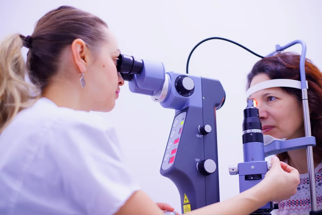
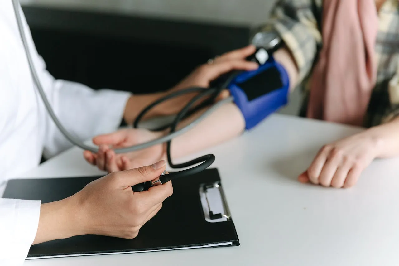
- Perform a physical examination. Your doctor will measure your blood pressure. You will also have an eye exam done. A healthcare professional will apply special eye drops that widen your pupils. Thereafter, a special object with a magnifying lens, called a fundoscope (ophthalmoscope), will be used to look for signs of hypertensive retinopathy at the back of each eye.
- Perform eye imaging tests. Other tests that can be done to check for problems with your retina include:
- Fluorescein angiography. In this test, a special dye called fluorescein is injected into one of your veins. Then, a special camera is used to take images that can be used to check blood flow to your retina.
- Optical Coherence Tomography (OCT). This is a quicker test that uses light waves to take pictures of your retina.
What signs of hypertensive retinopathy might a doctor see?
On an eye exam and imaging test, the signs of hypertensive retinopathy that may be observed by the eye doctor include:
- Microaneurysms—enlarged small blood vessels which appear as tiny red dots
- Narrowing of tiny arteries (arterioles)
- Thickening of the arterioles’ walls
- Cotton wool spots—fluffy, white areas that resemble clouds
- Hard exudates—yellow plaques made of proteins and fats from leaky blood vessels
- Bleeding (hemorrhage) within the retina
- Papilledema—swelling at the point where the optic nerve leaves the eye
- Retinal blood vessels looking like copper wires or silver wires. They normally appear red.
- Arteriovenous (AV) nicking. Where a small vein (venule) is indented by a crossing arteriole.
What are the stages of hypertensive retinopathy?
Hypertensive retinopathy can be graded depending on how severe the damage in the retina is. Using different classification systems, signs of more serious damage are placed in higher stages. Examples of classification systems are:
Keith Wagener Barker (KWB) classification
This system has 4 grades, which include:
- Grade 1: Here, there is slight narrowing of the retinal arterioles.
- Grade 2: Grade 1 plus AV nicking and further narrowing of retinal arterioles.
- Grade 3: Grade 2 plus retinal hemorrhage, cotton wool spots and hard exudates.
- Grade 4: Grade 3 plus papilledema.
Sheie classification
This system has 2 components. One of them is the staging of hypertensive retinopathy and is similar to the KWB grades, with just an additional Grade 0, where the retina has no abnormalities.
The other component is the grading of arteriosclerosis (the abnormal thickening or hardening of the walls of arteries). The stages in this component are:
- Stage 0: No retinal abnormalities.
- Stage 1: Widening of the arteriole light reflex, where there is more light reflected at the center of the retina.
- Stage 2: Stage 1 plus arteriovenous crossing (arterioles cross over with venules).
- Stage 3: Copper wiring of arterioles.
- Stage 4: Silver wiring of arterioles.
Wong and Mitchell classification
According to this simpler three-grade system, hypertensive retinopathy may be:
- Mild: If one or more of arteriolar narrowing, arteriovenous nicking, and copper wiring of arterioles are present.
- Moderate: If one or more of retinal hemorrhage, microaneurysms, cotton wool spots, and hard exudates are present.
- Severe: If signs from moderate retinopathy plus swelling of the optic disc is present.
What are the complications of hypertensive retinopathy?
When hypertensive retinopathy is not treated, it may worsen and cause:
- Lack of blood flow to the optic nerve
- Blockage in the artery that supplies the retina (retinal artery occlusion)
- Blockage in the vein that supplies the retina (retinal vein occlusion)
- Macular edema (swelling in the center of the retina)
- Worsening of diabetes-associated retinopathy
- Retinal detachment (pulling away of the retina from its supporting tissues)
How is hypertensive retinopathy treated?
Treatment for hypertensive retinopathy focuses on controlling your blood pressure with antihypertensive medicines and lifestyle changes. Lowering your blood pressure can help your retinas recover over time. However, this may not be possible if retinal damage is severe.
Your doctor will prescribe appropriate doses of medications to reduce your blood pressure slowly. Reducing blood pressure very fast can worsen hypertension-associated damage in other organs. Examples of commonly used medicines include:
- Angiotensin-converting enzyme (ACE) inhibitors
- Calcium channel blockers
- Diuretics
Lifestyle changes that may help to control your high blood pressure include:
- Losing weight if you are overweight or obese
- Engaging in moderate-intensity exercise for at least 150 minutes weekly
- Limiting alcohol intake
- Stopping smoking
- Reducing salt intake
How can I prevent hypertensive retinopathy?
You can prevent hypertensive retinopathy by following the same lifestyle changes that are recommended for people with the condition. A healthcare professional may recommend that you lose weight, follow a specific diet plan, or reduce how much alcohol you drink.
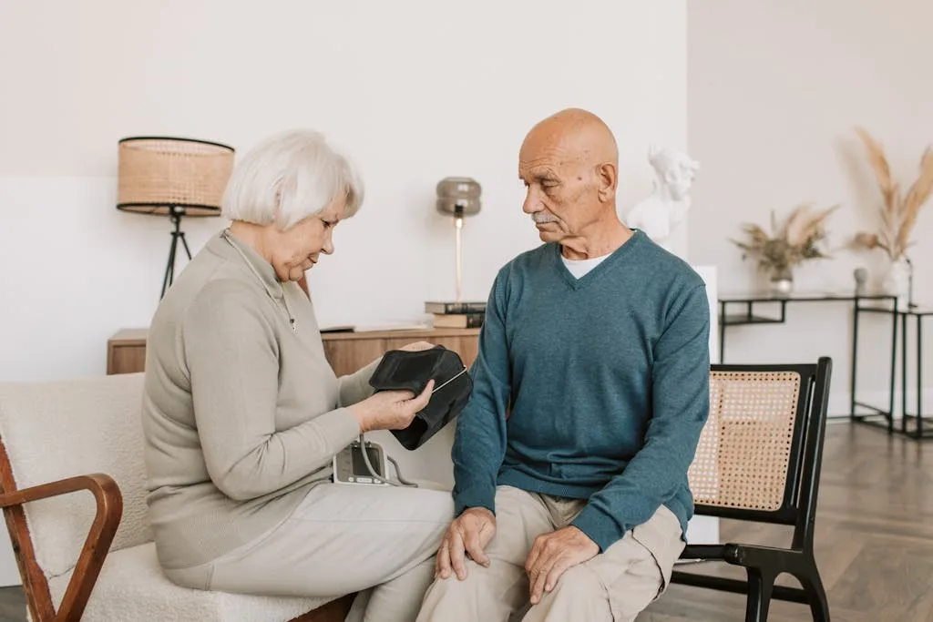
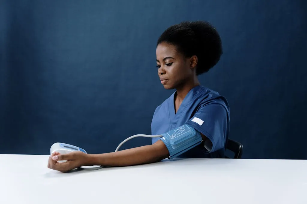
It is also important for you to have your blood pressure checked regularly. Being diagnosed with high blood pressure and having it treated early enough may prevent it from damaging your eyes and other organs.
Even when you have hypertension, you will benefit from regularly monitoring your blood pressure and having your eyes checked as often as your doctor suggests.
What should I remember?
Hypertensive retinopathy is a serious eye condition caused by high blood pressure. When blood pressure stays elevated, it can damage the delicate blood vessels in the retina, affecting vision. The longer hypertension goes untreated, the greater the risk of permanent damage.
Regular blood pressure checks and eye exams are essential for people with high blood pressure. Early detection of hypertensive retinopathy can help prevent further vision loss. Managing blood pressure through medication, diet, exercise, and lifestyle changes is key to protecting eye health. Taking these steps can significantly reduce the risk of vision problems and improve overall health.

